The first ray of the foot includes the first metatarsal bone and the base of the great toe (hallux). The first ray is a structurally significant part of the foot that plays a major role in its biomechanics and is required to assume specific postures to achieve balance during gait and to transfer weight during activities like walking and running. For this reason, it is critical to the overall health of the foot and understanding its normal anatomical position and alignment.
A plantarflexed first ray is a deformity of the foot that involves a tilt of the first ray downward. A plantarflexed first ray can be present at birth, also known as congenital. The child may be born with a deformity of the foot in which some or all of the toes are tilted downward. An acquired plantarflexed first ray involves changes of muscles and tendons around the first ray that occur over time due to injury, arthritis or other disease process. Finally, a repetitive stress injury such as running can cause a plantarflexed first ray over time.
Analyzing the plantarflexed first ray is very important when analyzing a patient’s foot function and their respective gait. Looking at a patient’s first ray during walking/running will give the practitioner an understanding of how forces are transmitted to the foot and how the patient might be gaining balance/stability. Looking further into this abnormality, research has proven that the plantarflexed first ray can cause the body to put increased amounts of stress on other areas of the foot. Pain can be a major result from the plantarflexed first ray. As Christensen and Jennings (2009) proved in their study, a plantarflexed first ray can even alter the alignment of the knee and/or hip, creating uneven stress through the lower extremities during movement.
For the patient with a plantarflexed first ray, both the physician and patient must understand the condition in order to best determine a course of treatment. Depending on the degree of severity, treatment can range from conservative to surgical, including the use of custom orthotics to treat and alleviate discomfort, as well as physical therapy to strengthen and improve function of the affected muscles and overall foot. A patient with a plantarflexed first ray will exhibit changes in body motion as he or she walks. Specifically, the way in which a person with this condition transfers weight onto the foot will be different than that of a person with a normally aligned first ray. Typically, the heel hits the ground first during gait, followed by a smooth transfer of weight to the forefoot as the person continues to step forward. The weight-bearing forces are transferred from heel to ball in an even distribution across the plantar surface of the foot. In contrast, the individual with a plantarflexed first ray transfers an uneven amount of weight onto the foot, pushing more weight onto the outside of the foot. This alteration in biomechanics can cause the individual to develop abnormal compensations which can have further effects on other joints of the legs, as well as the hips and lower back.
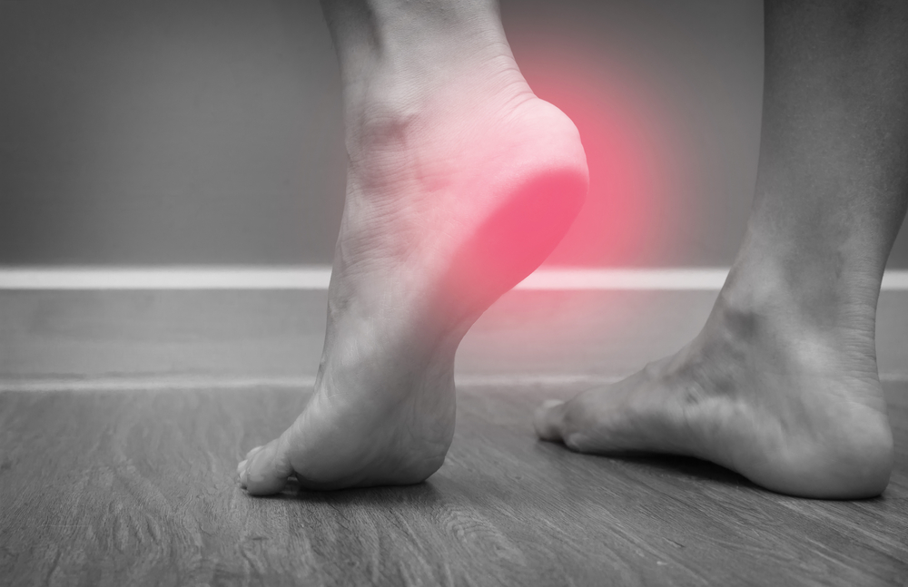
A plantarflexed first ray is a pathology that can cause a host of clinical problems, particularly metatarsal pain. As the first ray becomes plantarflexed, other parts of the foot must compensate and endure increased stress, creating pain in the forefoot, as well as the midfoot and even the heel. People with this deformity may even alter their gait, leading to an increased risk of a fall or ankle rollover, placing other joints of the lower limb at risk for tendonitis or other forms of arthritis. The patients with a plantarflexed first ray are at greater risk for injuries of the foot and lower limb of various origins.
A plantarflexed first ray is commonly associated with other deformities of the foot. The cavovarus foot deformity is a typical example. Characterized by an excessively high arch and possible inversion of the foot, cavovarus feet may cause individuals to experience difficulties with mobility and stability. The implications of a first ray that is positioned abnormally may add to these difficulties, especially during the toe-off and push-off portions of gait. As a result, identifying and understanding first ray function is an important aspect of evaluating and treating individuals with cavovarus feet (Krähenbühl and Weinberg, 2019).
Implications of a plantarflexed first ray can be addressed through clinical treatment of the associated biomechanical defects and pain. Treatment options may include orthotic devices to support the foot and redistribute weight to alleviate pain on the bottom of the foot (Van Beek and Greisberg, 2011). Additionally, physical therapy can be prescribed to treat muscle weakness around the foot. In severe cases, with disabling symptoms and poor function, surgical correction may be an option. Understanding the implications of a plantarflexed first ray is important for management of the patient with this deformity and for overall foot health and function. The treatment options for a plantarflexed first ray in the foot vary depending on the degree of deformity and the complaints of the patient.
Initial conservative management begins with the use of foot orthotics. Functional foot orthotics are placed into a well-fitting flat shoe. The aim is to support the first ray into a more neutral position. Research has shown orthotics to be effective in treating this condition, improving pain and function (Ballesteros-Mora et al., 2025; Mojica and Early, 2017). Patients may find additional support from shoes with a wider toe box. The type of orthotic chosen will be dependent on various factors including foot shape, and how a patient walks. It is essential to obtain correct foot orthotics to allow for optimal gait.
When conservative management fails to alleviate pain or when there is marked limitation of function, surgical options may need to be considered. The indications for surgery are based on several parameters and can include the severity of the deformity, the amount of pain the patient has, as well as how much the condition impacts the patient’s activities of daily living. The literature has well-described both osteotomies to correct the position of the first ray as well as soft tissue procedures that aim to stabilize the foot externally (Hild and McKee, 2011; Martin et al., 2012). Typically the patient will have been managed with multiple non-surgical treatments prior to consideration for surgical intervention. Each patient must be evaluated on an individual basis and surgery should be considered carefully for any indication.
Management of a plantarflexed first ray involves an interdisciplinary approach. Orthotic management and possible surgical intervention are most effective when supplemented by and coordinated with a physical therapy program focused on enhancing muscle strength and foot stability as well as education on the foot biomechanics. Patients can perform home exercises and benefit from exercises conducted in the clinic. This is supplemented by an educational program teaching patients on appropriate shoe gear selection, walking techniques, and other interventions that promote optimal foot function. (Christensen and Jennings, 2009).
Management of the plantarflexed first ray includes a thoughtful combination of conservative and surgical interventions. Knowledge and management of the plantarflexed first ray includes a life long process of patient education and management, as well as aggressive physical therapy and strengthening. The end result can be quite good for these patients.
Citations:
Hild, G.A. and McKee, P.J., 2011. Evaluation and biomechanics of the first ray in the patient with limited motion. Clinics in podiatric medicine and surgery, 28(2), pp.245-267. https://www.podiatric.theclinics.com/article/S0891-8422(11)00009-7/abstract
Christensen, J.C. and Jennings, M.M., 2009. Normal and abnormal function of the first ray. Clinics in podiatric medicine and surgery, 26(3), pp.355-371. https://www.podiatric.theclinics.com/article/S0891-8422(09)00011-1/abstract
Morgan, O.J., 2023. Biomechanics of First Ray Hypermobility (Doctoral dissertation, Anglia Ruskin Research Online (ARRO)). https://aru.figshare.com/articles/thesis/Biomechanics_of_First_Ray_Hypermobility/23767392
Ballesteros-Mora, M., Munuera-Martínez, P.V., Tovaruela-Carrión, N., Sáez-Díaz, A. and Ramos-Ortega, J., 2025. Study of Force Changes Based on Orthotic Elements Under the First Ray. Applied Sciences, 15(14), p.7708. https://www.mdpi.com/2076-3417/15/14/7708
Schuberth, J.M. and Babu-Spencer, N., 2009. The impact of the first ray in the cavovarus foot. Clinics in Podiatric Medicine and Surgery, 26(3), pp.385-393. https://www.podiatric.theclinics.com/article/S0891-8422(09)00033-0/abstract
Martin, H., Bahlke, U., Dietze, A., Zschorlich, V., Schmitz, K.P. and Mittlmeier, T., 2012. Investigation of first ray mobility during gait by kinematic fluoroscopic imaging-a novel method. BMC musculoskeletal disorders, 13(1), p.14. https://link.springer.com/article/10.1186/1471-2474-13-14
Krähenbühl, N. and Weinberg, M.W., 2019. Anatomy and biomechanics of cavovarus deformity. Foot and ankle clinics, 24(2), pp.173-181. https://www.foot.theclinics.com/article/S1083-7515(19)30014-2/abstract
Colò, G., Fusini, F., Marcolli, D., Leigheb, M. and Surace, M.F., 2025. Hindfoot Valgus and First Ray Insufficiency: Is There Correlation?. Surgeries, 6(2), p.26. https://www.mdpi.com/2673-4095/6/2/26
Mojica, M.N. and Early, J.S., 2017. Foot Biomechanics. Atlas of Orthoses and Assistive Devices E-Book, p.216. https://books.google.com/books?hl=en&lr=&id=6RNBDwAAQBAJ&oi=fnd&pg=PA216&dq=plantarflexed+first+ray+foot+biomechanics&ots=ryYwq7mAD1&sig=PTLbqUWnsse2EU4M-Cw_TvGEHAA
Van Beek, C. and Greisberg, J., 2011. Mobility of the first ray. Foot & ankle international, 32(9), pp.917-922. https://journals.sagepub.com/doi/abs/10.3113/fai.2011.0917
University lecturer, runner, cynic, researcher, skeptic, forum admin, woo basher, clinician, rabble-rouser, blogger, dad.